What Is a Tissue Microscope?
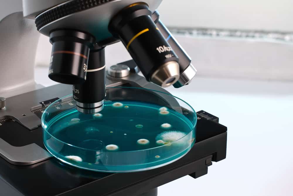
A tissue microscope is exactly what you would expect it to be: a specifically designed and nuanced microscope for analyzing live cells. Unlike other models, the tissue microscope has a small petri dish to contain the sample. Tissue microscopes typically feature an excellent magnification strength to view tiny cells.
The tissue microscope is designed to keep samples alive; therefore, a Petri dish is used rather than the sample slide for analysis. Unlike other models, the tissue microscope is inverted: the light source is directed from above rather than below. Despite being used for very detailed research, this type of microscope is incredibly user-friendly and easy to operate.
Features and Benefits of a Tissue Microscope
High-resolution imaging is essential for the detailed analysis of tissue cells, and the tissue microscope is well-equipped for this specific purpose. As the model is so user-friendly, the user can process images much faster and multiple samples very quickly, which is a huge benefit when processing a large amount of material.
Live imaging is essential in microbiology to observe the development of specimens, otherwise known as in vivo, a scientific term derived from the root Latin term meaning “within the living.” The tissue microscope is perfectly designed with live imaging capabilities to capture the in-process assessment of samples.
Tissue samples are often translucent and difficult to analyze, but the tissue microscope’s contrast capabilities are available to assist with this. The light source is applied from above; this technique is for increased dimensioning of the sample under review.
Some advanced imaging options available with the tissue microscope allow for a greater analysis of cell cultures and provide a multi-dimensional view. The multispectral and multiphoton capabilities for this type of microscope provide the ability to observe more closely and extract essential information.
Tissue Under the Microscope
All tissues in the body are one of four distinct types of tissue: nervous, epithelial, muscle, and connective tissue. Each of these four types appears in numerous places throughout the body and behaves differently under the microscope.
- Nervous tissue is composed of two types of cells: neurons and glia. These cells support the nervous system via communication and computation with the brain. Nervous tissue under microscope has an almost purple star-like appearance.
- People often describe adipose tissue under microscope as having a spongy-lung-like appearance when magnified, and it is easy to see why. When prepared for a sample and scrutinized under magnification, the cells appear wide and empty, which they are – the space is used to house fat droplets.
- Connective tissue has an essential job in the body – to bind and hold together the area around vital organs and other tissues. It comprises three main elements: cells, protein fibers, and ground substance. Due to its nature, connective tissue under microscope appears loose and fibrous.
- Bone tissue has a critical job within the body – to provide strength and stability to the skeletal frame and consists of both compact and cancellous tissue. Bone tissue microscope images are interesting because they are two separate-looking tissue components.
- Reticular tissue is loose and fibrous connective tissue. It predominantly serves to scaffold other tissue and protect organs. Reticular tissue under microscope is loose and mesh-like.
- Areolar tissue under microscope is interesting to look at and is very distinctive since it lacks organization and has the appearance of multiple layers of thread or a spider’s web. The tissue serves as a cushion to vital organs and also disperses nutrients.
- Muscle tissue under microscope looks different depending on the type. There are three main types of muscle tissue: cardiac, smooth, and skeletal. Both skeletal and cardiac muscle tissue appears to be striped, also called striated, whereas smooth muscle tissue is homogenous and non-striated.
- Epithelial tissue lines the area surrounding the vital organs and blood vessels. Epithelial tissue microscope images have a sheet-like appearance.
Applications of Tissue Microscopy
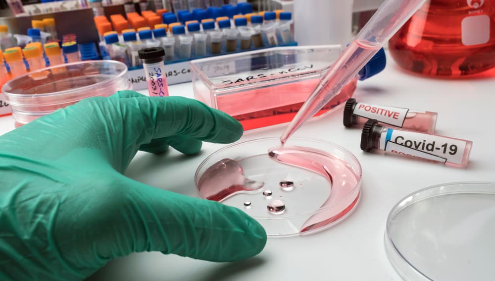
Using tissue microscopy in research and development is integral to significant medical breakthroughs and discoveries. A very recent example of this was throughout the COVID-19 pandemic. Researchers could quickly identify the virus, its traits, its behavior, and how it mutated through tissue microscopy.
In biological and life science research, tissue microscopy resulted in a recent breakthrough regarding de-crowding molecules to analyze previously inaccessible imaging of new cellular structures. Findings were published from MIT in August 2022.
In pharmaceutical and drug development, close analysis of tissue types is imperative in understanding cellular and diseased bio-markers to tailor specific treatments and drugs.
FAQs
How is tissue microscopy different from other types of microscopes?
The unique feature of a tissue microscope is that you aim to keep your specimen or subject alive throughout the evaluation. The Petri dish element of this, rather than a preserving slide, sets this microscope apart. Similarly, another interesting feature of this microscope is the inverted scope, where the lighting is actually placed above the subject rather than below and shining up, illuminating the subject.
Can a tissue microscope be used for animal studies?
Yes, all tissue microscopes can be used to study animal tissue.
What are the advantages of live imaging?
Live imaging is a powerful tool, and there are a few main benefits:
- Observation of cellular developments as they occur
- The ability to keep samples in a native environment, thus leading to more natural results
- Interactions between cells can be closely observed
What are the most common contrast techniques?
Frits Zernike won the Nobel Prize in Physics for inventing the phase contrast microscope. He recognized the challenges and complexity of analyzing translucent cells, and to mitigate this, he invented a microscope that could provide contrast to the specimens under evaluation.
Now there are multiple methods of creating contrast. Modern common and popular techniques include dark field, fluorescence, polarization, and phase contrast.

