It is incredible how far technology has come, especially in research and education. Thanks to advanced engineering, a wide range of microscopes is available for all types of work in various industries and fields. Today’s topic: electron microscopes.
While most people know a lot about using light microscopes, typically found sitting on students’ desks in high school and college, there is another popular option capable of much more.
Electron microscopy was developed in 1931 and has grown and developed exponentially over the last 90+ years. It offers a scientific instrument to the world that can magnify objects up to 10,000,000x.
So what is electron microscopy? How does it work? Where can you use it? We will answer these questions and so many more.
How Do an Electron Microscopes Work?
The extremely high resolution of electron microscopes separates these instruments from the others. Instead of the typical beam of light to illuminate objects, an electron microscope uses beams of accelerated electrons.
The lens’s focus is fixed on an optimal microscope, and the distance between specimens varies. With an electron microscope, the distance between the specimen and the microscope stays relatively consistent. At the same time, there are variable focus lenses (zoom lenses), changing the focal length for more convenient views.
With up to 100,000 times shorter wavelengths, the electron microscope provides a clearer, more detailed illumination of small object structures. These objects can be much smaller than those seen with traditional microscopes.
Electron Microscopes: Parts and Functions
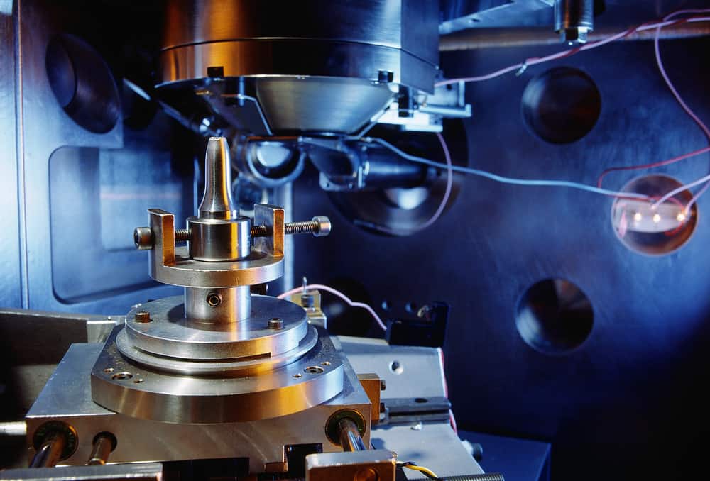
An electron microscope comprises four main parts with various accessories to improve its functions. These main parts include the electric gun, electromagnetic coils, microscope column, and fluorescent screen.
- Electron Gun: This is where the electron beam comes from. It is situated on top of the microscope and made from tungsten surrounded by a shield with a negative bias. This gun has an aperture, where the light travels from.
- Electromagnetic Coils: These electric wiring coils (or lenses) are set inside a metallic cylinder hollowed in the center. As the electric current passes through the coils, the magnetic field is the magnification lens.
- Microscope (Central) Column: This column is a form of protection for the operator. It is made from an evacuated metal tube. This tube keeps the X-rays generated from electrons striking the metal tube surface from harming the person using the microscope.
- Fluorescent Screen: This screen forms an image of the magnified object on a television. The screen is coated with chemicals that react excitedly, creating the image.
Some other features and accessories include:
- High voltage transformers
- Water cooling systems
- Filter systems
- Circulation pumps
- Vacuum pumps
- Refrigeration plants
Combining all these parts creates a massive microscope that forms images of target objects by scattering, dispersing, and projecting electrons onto a fluorescent screen.
Advantages of Electron Microscopes
What are the advantages of an electron microscope? Well, there are many, along with a few disadvantages as well. Let’s take a look.
| Advantages | Disadvantages |
| 500,000x magnification Higher resolution compared to a light microscope Versatile – can be used with organic and inorganic specimens Compatible with a vast library of applications and other technology for broader use | Requires specific housing for use and maintenance Much larger than a typical light microscope Much more expensive than a light microscope Specimen needs extreme care and storage in a vacuum chamber |
Scanning Electron Microscopes
A scanning electron microscope uses the same process as a regular electron microscope but is used for scanning surfaces. This item is used in various industries, including but not limited to forensic science, fabrication, medicine, etc.
What Does a Scanning Electron Microscope Do?
An SEM or scanning electron microscope, like a transmission electron microscope, uses tungsten filament lamps that contain electromagnetic lenses and electrons stationed at the top of the column.
As thermal energy is applied to an electron object, the electrons are emitted and move rapidly toward the opening or anode with a positive charge. The beam that passes through the anode then activates the scattering of primary and secondary electrons off the object’s surface.
The interaction between the beams and the targeted specimens will produce a signal that provides essential information and details regarding the surface composition and topography.
Once complete, the object must undergo a fixation, dehydrating, and drying process to prevent the cell’s features from collapsing when exposed to the microscope’s vacuum.
Parts of Scanning Electron Microscope
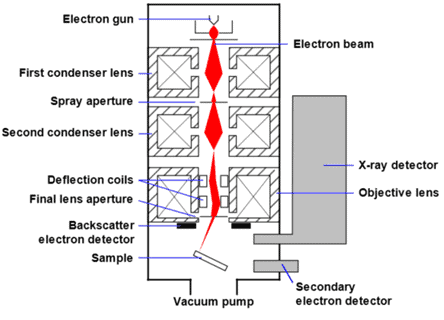
For a better understanding of how scanning electron microscopes work, take a look at each part and its essential role in the instrument’s inner workings.
- Electron gun
- First condenser lens
- Spray aperture
- Second condenser lens
- Deflection coils
- Final lens aperture
- Backscatter electron detector
- Sample chamber
- Vacuum pump
- Secondary electron detector
- Objective lens
- X-ray detector
From the top of the column, electrons are rushed downward toward the multiple lenses and apertures, creating a laser-focused electron beam. This beam strikes the sample’s surface, then scans it with the help of coils located directly above its objective lens.
After the scanning is complete, the signals are passed to the detectors, which then collect the data and recreate the image you will eventually see on the screen.
While many parts are similar to the basic electron microscope, there are additional parts, making it more advanced, larger, and capable of conducting different tasks.
The scanning electron microscope magnification and depth of field are much higher than that of optical microscopes, providing an exact copy of the surface of the specimen.
Advantages of a Scanning Electron Microscope
Scanning electron microscopes are beneficial in various industries, providing a tool that can accomplish many jobs no other instrument can. However, along with its advantages, it also has a few disadvantages.
| Advantages | Disadvantages |
| Easy learning curve Can quickly analyze surfaces of solid objects Can generate digital data for sharing and transport Gets essential information within five minutes | Larger than other microscopes, hard to transport Extremely expensive Requires frequent maintenance (consistent voltage and cooling systems) |
The cost of scanning electron microscopes is the most significant disadvantage. These scientific instruments cost anywhere between $50,000 to $200,000. This cost will vary depending on your desired type and its configurations, resolution, components, etc.
Another disadvantage noted is the large size of the unit, making it hard to transport. This issue can be rectified by purchasing a portable scanning electron microscope, which is more compact.
Field Emission Scanning Electron Microscope
A specific type of scanning electron microscope is an FE-SEM or field emission scanning electron microscope. These microscopes work with electrons rather than light, creating microstructure images of objects through emission field sources.
Field emission scanning produces a clearer image than the conventional scanning electron microscope. It provides better spatial resolution and less distortion, creating 3-6 times better images.
This instrument uses a field emission cathode inside the scanning electron microscope gun. This cathode creates a narrower probing beam than the regular scanning microscope, with low and high electron energy. This produces minimal sample damage and improved spatial resolution.
How Much Does a Field Emission Scanning Electron Microscope Cost?
Similar to our answer for the other microscopes in this article, the cost of a field emission scanning electron microscope will vary depending on your type and the features you are looking for.
As you can imagine, these microscopes are very costly due to the advanced technology and equipment used to design them. The most expensive field emission scanning electron microscope on the market today costs $10,000,000. However, the average price will range between $125,000 to $900,000. Definitely not a cheap tool, but worth every penny.
Advantages of a Field Emission Scanning Electron Microscope
A field emission scanning electron microscope is crucial in various industries, mainly used to study semiconductors and metals. You will also find these microscopes have been used to obtain a few images of biological specimens.
There are numerous advantages to having a field emission scanning electron microscope in these fields.
| Benefits of FESEM |
| Examine tiny areas with clear and precise images Reduces the chances of damage to the image High-quality images with low voltage Do not have to coat samples ahead of time |
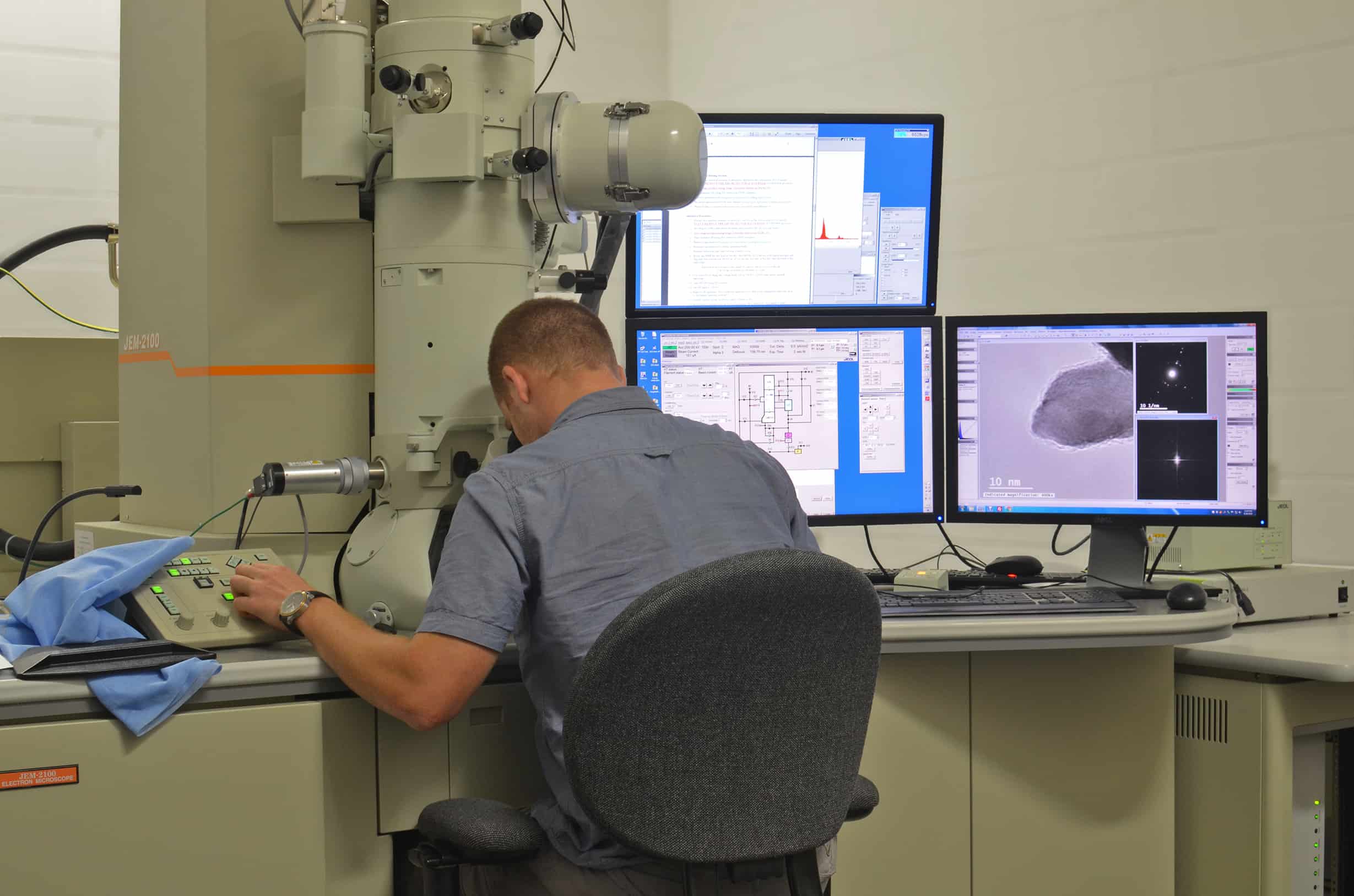
Transmission Electron Microscopes
A transmission electron microscope applies the same techniques as the other two instruments by utilizing electron beams for illumination rather than lighting. However, this microscope creates an image as the electrons move through the specimen instead of scanning it. The image is sent to a digital recording system, allowing you to view, focus, and record your data.
This technology allows the user to see tiny objects, far too small for the human eye, in great detail.
How Does a Transmission Electron Microscope Work?
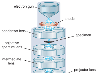
To better understand how a transmission electron microscope works, you must know all the components used to assemble the instrument, creating three specific and crucial systems.
- Electron Gun
- Electron Beam
- Condenser System
The electron gun is part of the microscope that produces electrons and focuses, controls, and deflects the beam. The cathode covered with oxide is heated by a coil that emits electrons. These electrons then get pulled toward the first anode, a positively charged sleeve.
As the beam moves along, it is contoured, then electrostatically collimated or condensed and forced through a metal disk with a hole, finally striking a screen coated in phosphor.
As the electrons move through the system, the alignment of the electron gun and control over it is critical to the overall outcome of the task. One wrong move or measurement, and you will have to start the process all over.
Once the electrons pass through the object, it develops a grid that travels toward a specimen stage. It moves through a projector lens and magnifies the image, making it easy to see in sharp, clear, detailed focus.
Advantages of a Transmission Electron Microscope
Using a transmission electron microscope has many benefits. However, there are a few disadvantages to them as well.
| Advantages | Disadvantages |
| Has the highest magnification abilities (up to 1,000,000x or higher) Offers detailed information on compound and element structures Produces high-quality images with accurate details Easy to learn and operate Compatible with various applications in various fields | Takes a lot of time to prepare Requires special training to operate and analyze specimens Only specimens that are electron transparent can be used Specimen has to be very small Images only come in black and white |
How Much Does a Transmission Electron Microscope Cost?
Various manufacturers produce transmission electron microscopes, each selling their products at an extremely high price, with the average cost is around $100,000.
Atom Electron Microscope

An atom electron microscope is an advanced electron microscope that achieves exceedingly high resolution using electron beams, as opposed to light, to illuminate the specimen (atoms). This work is referred to as atom microscopy.
This task is no easy feat, as atoms are an element’s tiniest unit. This unit retains the same elements’ properties even when broken down. This means its protons, neutrons, and electrons will not change.
Many typical electron microscopes can be used to see atoms since they are built to magnify objects more than 500,000x.
A transmission electron microscope is the best option for detecting atoms and nanoparticles.
Light Microscopes vs. Electron Microscopes

There are two main types of microscopes out there, including the light microscope and the electron microscope. While each offers significant benefits to the user, they differ in many ways.
The most obvious difference is in illumination. When using a light microscope to inspect a specimen, you will need a light source such as a halogen bulb or LED. In contrast, the electron microscope doesn’t require a light source. These products use electrons for illumination.
Another glaring difference between the two is the maximum magnification they provide. The light microscope is ideal for the study of internal structures. Place a sample on a slide and push it underneath the lens and lighting. The glass lens amplifies or magnifies the object, allowing you to see it in greater detail.
However, the magnification is only enough to see specific details in specific specimens, live or dead, and can only reach a maximum magnification of 1000x. This is much lower than an electron microscope’s maximum magnification of 2,000,000x.
Electron microscopes are often used to study external surfaces such as small organisms and cell structures. You can use these instruments on dead or dried specimens, but they will not work on live ones.
If you have ever used a microscope at school or work, you probably noticed that the specimen is always seen in 2D. This can be helpful when looking at organisms like cells or bacteria. The user can see a specimen in a full 3D view in electron microscopes.
| Feature | Light Microscope | Electron Microscope |
| Maximum Magnification | 1000x | 2000000x + |
| Source of illumination | Light | Electrons |
| Images | Colored | Black and white |
| Field of Use | Internal structures | External structures |
| Lenses | Glass | Electromagnetic |
| Dimensional view | 2D | 2D and 3D |
Which Microscope Should You Use?
You should determine the type of microscope you need based on the results you hope to see. You can use a light microscope to examine objects like tissues and cells in places like biology class to learn about algae, bacteria, fungi, or plant and animal cells.
You would use an electron microscope to see tiny specimens you can’t even view with a light microscope. You can examine the composition and structure of an atom under an electron microscope as well as organelles, tissue, and molecules. You can also use an electron microscope to examine metals, circuits, semiconductors, etc.
In short, light microscopes are used to do less invasive examinations that don’t require a high level of magnification. An electron microscope is used in intricate situations where a higher level of technology, accuracy, and magnification is needed.
Final Thoughts
The world of microscopes is vast and can get confusing when deciding which specific option is best for each situation and required outcome. Before you purchase a microscope or try to learn how to use them for your work, take the time to research them for accurate and detailed information regarding each option.

