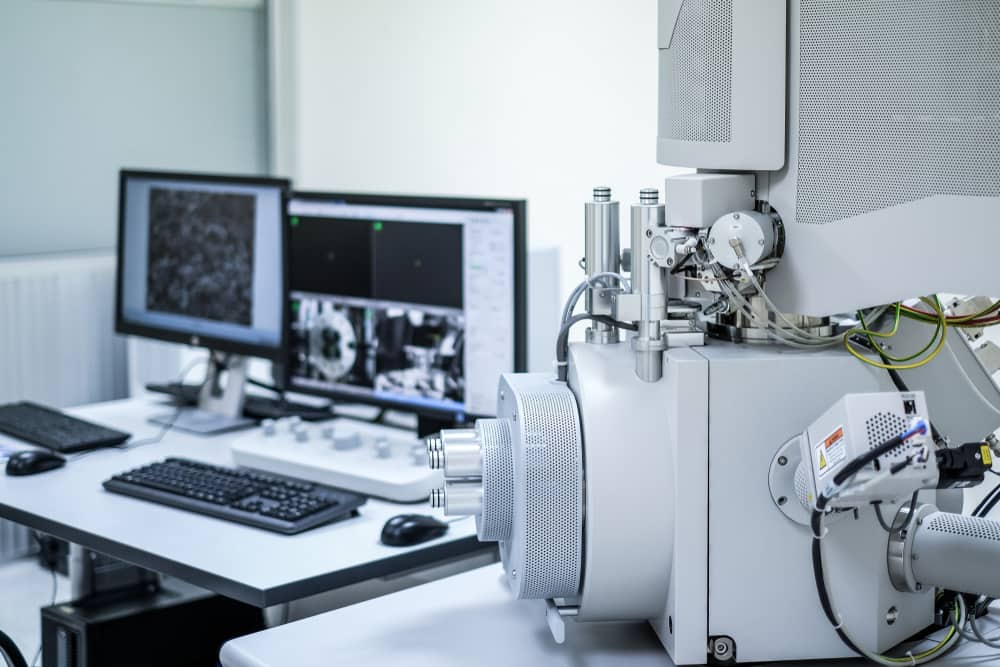
Confocal scanning microscopes are versatile tools with a ton of different scientific applications. If you work in scientific research, chances are you have encountered this type of microscope before and are aware of its capabilities.
These microscopes use powerful technology to scan specimens up close without damaging the sample. It can be beneficial for producing high-quality images and getting the information you need while examining a specimen.
Let’s look at what these microscopes are, how they work, and the different applications in which they can be applied.
Contents
What Is a Confocal Laser Scanning Microscope?
A confocal scanning microscope (CSM) is an optical microscope that uses a laser beam to scan a specimen, producing high-resolution images with excellent clarity and contrast.
Confocal microscopy uses a point-by-point scanning method to produce images of biological samples, which can then be reconstructed into 3D images. A confocal laser beam illuminates a small spot on the sample, and the reflected light is collected through a pinhole aperture. The pinhole aperture helps to block out-of-focus light, which is one of the primary sources of blur in traditional microscopy. This allows for producing highly detailed and clear images of the sample.
The collected light is then detected by a photomultiplier tube and converted into an electrical signal, which is then used to generate a digital image. Because the confocal laser is scanned across the sample, multiple images are taken from different angles, which can be combined to produce a 3D reconstruction of the sample.

When Are Confocal Scanning Microscopes Used?
As we touched on above, scientists utilize confocal scanning microscopes for a wide range of uses. However, four specific fields of science use them most often: biological research, materials science, medical imaging, and forensic science.
Let’s break down their uses in these spaces:
- Biological research: CSMs are widely used in biological research, particularly neuroscience, cell biology, and developmental biology. You can use them to visualize cellular and subcellular structures, including organelles, nuclei, and cytoskeletal elements, with high resolution and clarity. CSMs can also study live cells or tissues, allowing researchers to observe biological processes in real time.
- Materials science: CSMs can image materials and structures at the nanoscale. They are particularly useful for studying complex structures, such as porous or composite materials, where traditional microscopy techniques may not provide sufficient resolution or contrast.
- Medical imaging: CSMs can study tissue samples and diagnose diseases. You can use them to produce high-resolution images of cells and tissues, which can identify abnormalities or monitor disease progression.
- Forensic science: CSMs can be used to study trace evidence, such as hair or fibers, with high resolution and clarity. You can also use them to study fingerprints or other physical evidence, helping identify suspects or establish the chain of events in a crime.
Confocal scanning microscopes are a powerful tool for biological and medical research and other fields requiring high-resolution sample imaging.
The Benefits of Using Confocal Scanning Microscopes
There are several benefits of using confocal scanning microscopes for scientific research. Some of their perks include:
High resolution
Confocal scanning microscopes produce high-resolution images with excellent clarity and contrast. This allows for a detailed examination of the structures and functions of cells and tissues not found elsewhere. CSMs can identify three-dimensional images of cellular structures, such as organelles, nuclei, and cytoskeletal elements.
This is particularly useful for studying complex biological systems, such as the nervous system, where high-resolution imaging is critical for understanding the organization and function of neural circuits. This level of detail can help researchers better understand cellular processes and functions.
3D imaging
Confocal scanning microscopes allow for the reconstruction of 3D images of biological specimens. This provides researchers with a complete view of the studied sample and allows for examining structures that would otherwise be difficult or impossible to visualize with conventional microscopes.
Reduced background noise
Confocal scanning microscopes use a pinhole aperture to block out-of-focus light, which reduces background noise and improves image contrast. This is particularly useful for imaging thick specimens, where background noise can be a major issue.
Live cell imaging
Confocal laser scanning microscopes can be used for the live imaging of biological specimens, allowing researchers to observe dynamic processes such as cell division and migration in real time. This is particularly useful for studying cellular and developmental biology, drug discovery, and development.
Versatility
Confocal laser scanning microscopes can be used for various applications, including imaging living cells and tissues, histology, pathology, medical imaging, and material science. You can use them to investigate biological and material samples with high resolution and clarity, and their use is becoming increasingly common in research, medicine, and other areas.

In Conclusion
For scientific research, a scanning confocal microscope can be a powerful tool for examining materials at a cellular level. A CSM can provide a high-resolution, 3D image of biological specimens while reducing background noise. And, thanks to its live cell imaging capabilities, it is versatile across multiple scientific applications.
These microscopes are essential in biological research, materials science, medical imaging, forensic science, and many other fields. They allow researchers to visualize cellular and subcellular structures and to study complex materials and structures at the nanoscale with high resolution and clarity.
While CSMs are expensive and require specialized training, they are a valuable tool for investigating a wide range of biological and material samples with high resolution and clarity. It’s evident in their rising popularity in the scientific field, with their use becoming increasingly common in research, medicine, and other fields.

