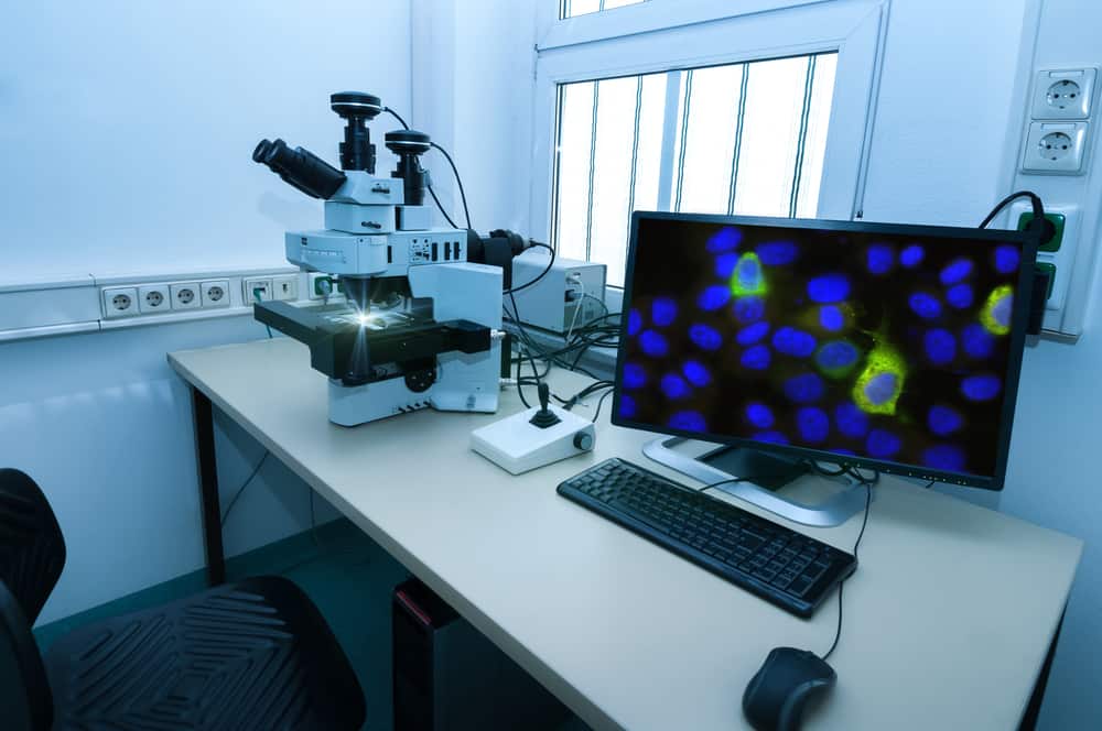
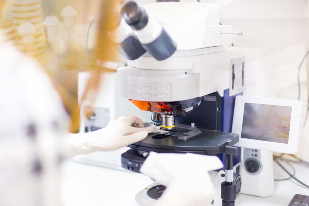
EVOS microscopes offer advanced images and analysis of cells (living or dead). The features and software that come with this tool provide photos and data you can’t get anywhere else.
If you want an innovative way to teach a class, conduct a detailed presentation, or publish your findings with sensational imagery, an EVOS microscope is for you.
With four EVOS models to choose from, you can get the perfect setup for your specific needs.
What Are EVOS Microscopes?
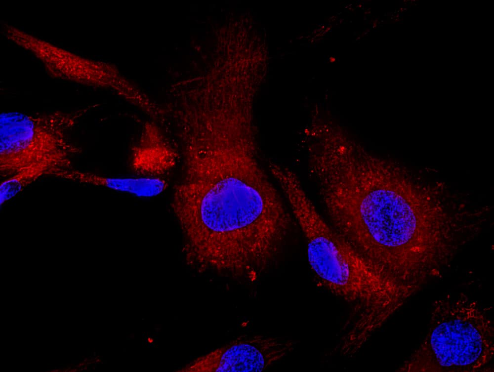
EVOS microscopes are high-quality image-capturing devices that can do the job of a traditional microscope but provide better imagery and still photos of the specimen (antibodies, microorganisms, etc.) you are looking at. Typically these microscopes use fluorescence microscopy lighting.
An EVOS microscope is the ideal instrument to use if you are a teacher, scientist, or preparing to conduct a presentation with incredible views for cell maintenance and tissue and cell cultures. It will automatically focus and adjust the brightness of your lighting while capturing detailed pictures and data in a very short amount of time.
Since the introduction of this intuitive microscope system software in 2009 by the America Microscope Group, many models have followed the first, with various applications depending on your needs.
Features of EVOS Microscopes
- High-resolution cameras
- Intuitive software
- Bright light sources with fluorescence microscopy
- User-friendly software
- Easy to set up
- Scans multiwell dishes at the same time
- Automatic or semi-automatic configurations
- Compact and easy to transport
4 EVOS Microscopes: Different Models for Your Specific Needs
Check out the top four EVOS microscope models today.
EVOS FLoid Imaging System
The FLoid imaging system is considered one of the easiest cell imaging systems available worldwide. It is exceptionally fast and straightforward to use. This system can capture three-color fluorescent, high-quality images in just one minute.
This EVOS fluorescence microscope is perfect for cell culture applications that need the help of fluorescence for visual quality or a fast and easy image of your cells.
Features
- Quick and easy installation: Installs on its own in about 10 minutes. No calibration, alignment, assembly, or maintenance is required.
- Quick images: Images are ready to go within 60 seconds. No warming up, cooling down, or filter changes are required.
- Basic fluorescence color: Three-color fluorescence. Offers focus assistance plus adjustable brightness, contrast, and focus.
- Multilingual: Seven-language interface.
- Blocks ambient light: Use on a benchtop, no longer required to stay in a darkroom when taking fluorescent cell images.
EVOS M7000 Imaging System
This EVOS microscope offers users the benefit of dual dedicated cameras. This includes fluorescent imaging and colorimetric imaging. It is a fast and reliable unit offering the EVOS microscope resolution known for quality and efficiency.
The EVOS M7000 imaging system is a fully automated imaging system that can quickly scan multiwell plates with live or dead cells, providing fast images with precise focus and a large data processor.
This microscope is the Ideal option for spatial gene expression research.
Features:
- EVOS onstage incubator option: Control humidity, gasses, and temperature for live cell analysis.
- High speed: Scans 3-fluorescence channel, 96-well plate within five minutes.
- Image stitching and tiling: Optimized light cubes for high-class images.
- Time-lapse imaging: Get accurate, clear images side by side from cell transformation over time.
- Single-click multichannel capture: Take quick, detailed pictures.
- Autofocus: Adjusts automatically to provide clear images.
- Z-stack capability: Provides clear images with various depths, thickening the plane of focus.
EVOS M5000 Imaging System
This EVOS inverted microscope system offers advanced image analysis and analysis with 3D analysis and visualization and 2D and 3D deconvolution. Thanks to the infinity-corrected optical system offering accurately detailed, precise imaging with high-quality light cubes and hard-coated filters, you can confidently use this microscope.
This system is ideal for research and not diagnostic procedures, making it the perfect choice for botanists, scientists, and educators.
You will get picture-perfect, detailed views of every specimen using this application with a high-resolution, four-color fluorescence camera, delivering pictures within seconds.
Features:
- Self-installation: Installs within minutes without needing alignment, calibration, or maintenance.
- Automated: Can count cells and take confluence measurements automatically.
- Advanced performance: Time-lapse imaging, z-stack capabilities, and single-click multichannel image capture.
- Four-color fluorescence: Uses advanced color options with four-color fluorescence, color images, and transmitted lighting.
- Biosafety cabinet and hood compatible: Use this system with a biosafety hood or cabinet.
- Advanced imaging: Performs 3D visualization and analysis and 2D and 3D deconvolution.
EVOS XL Core
The EVOS XL Core best suits bright field imaging cell culture applications. You will get high-quality, complex, detailed bright field cell images in one minute. This system requires a cell culture room or a hood, offering high-resolution and high-quality colored images with just a few clicks.
Features:
- Optional mechanical stage: Includes a fixed stage of more objectives based on needs.
- High-quality bright field color: Phase and bright field contrast images with the ability to adjust color tone, focus, and light intensity.
- Self-installation: Installs within two minutes without aligning or changing the bulb.
- Compatible with cabinet or hood: Use with a biosafety cabinet or hood.
- Adjustable speed: You choose the speed and resolution.
Choosing the Best EVOS Microscope

Here are some things to consider when choosing the best EVOS microscope.
EVOS Microscope Price
The price of the EVO microscope system you buy will vary depending on the model you choose and the accessories you get with it. You can purchase the microscope itself, or you can build an entire station. These setups can cost anywhere from $1000 (basic) to $50,000 (advanced).
EVOS Versatility
Do you want a laboratory workhorse with color and fluorescence imaging (EVOS M5000 and EVOS M7000) or a basic lab microscope to tackle cell culture applications (EVOS FLoid or EVOS XL Core)?
Intended Use
The features of each microscope vary widely. The only product with motorized encoded X/Y scanning is the EVOS M5000. All options have fluorescence channels except for the EVOS XL Core. Do you want your microscope to come with an associated printer? The only option you have in that regard is the EVOS FLoid. Take a close look at the features of each microscope before buying your own.
Other software attributes to consider
- Embedded analysis
- Networking capabilities
- Intuitive onboard software
- Integrated reagent selection guides
Final Thoughts on EVOS Microscopes
Each EVOS microscope listed above can significantly affect how biologists, researchers, and teachers work. Before choosing a model to add to your lab, consider the article above and decide which option best meets your imaging requirements.

