
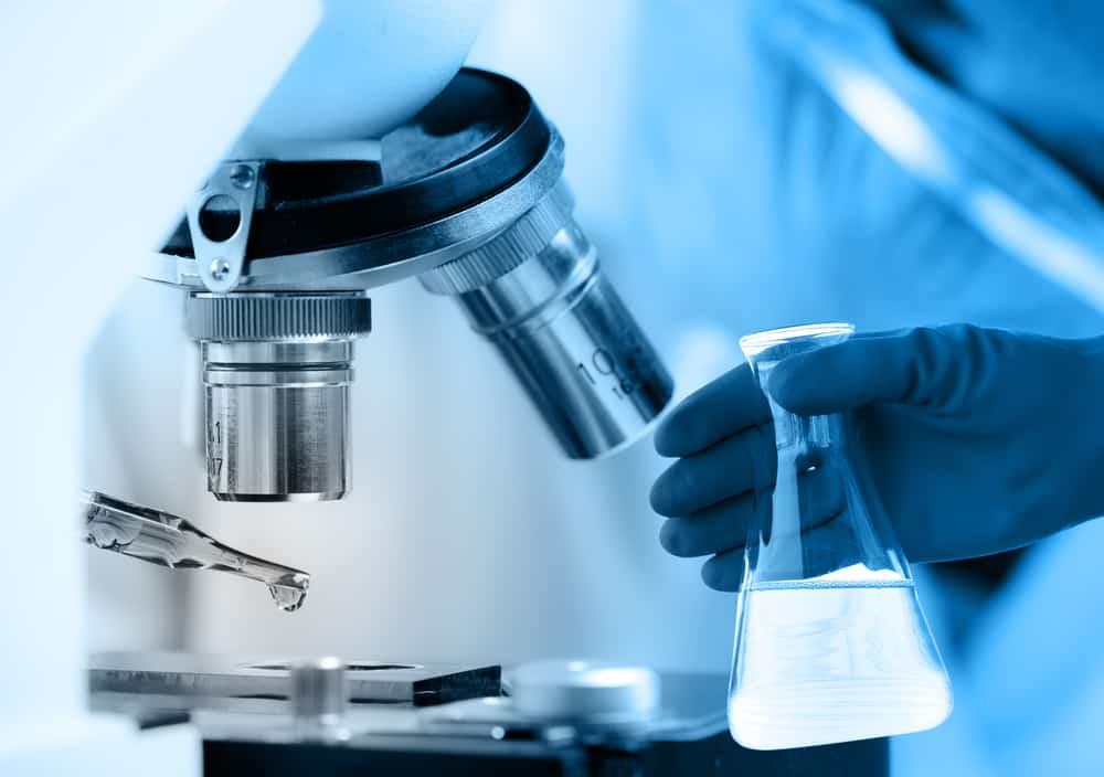
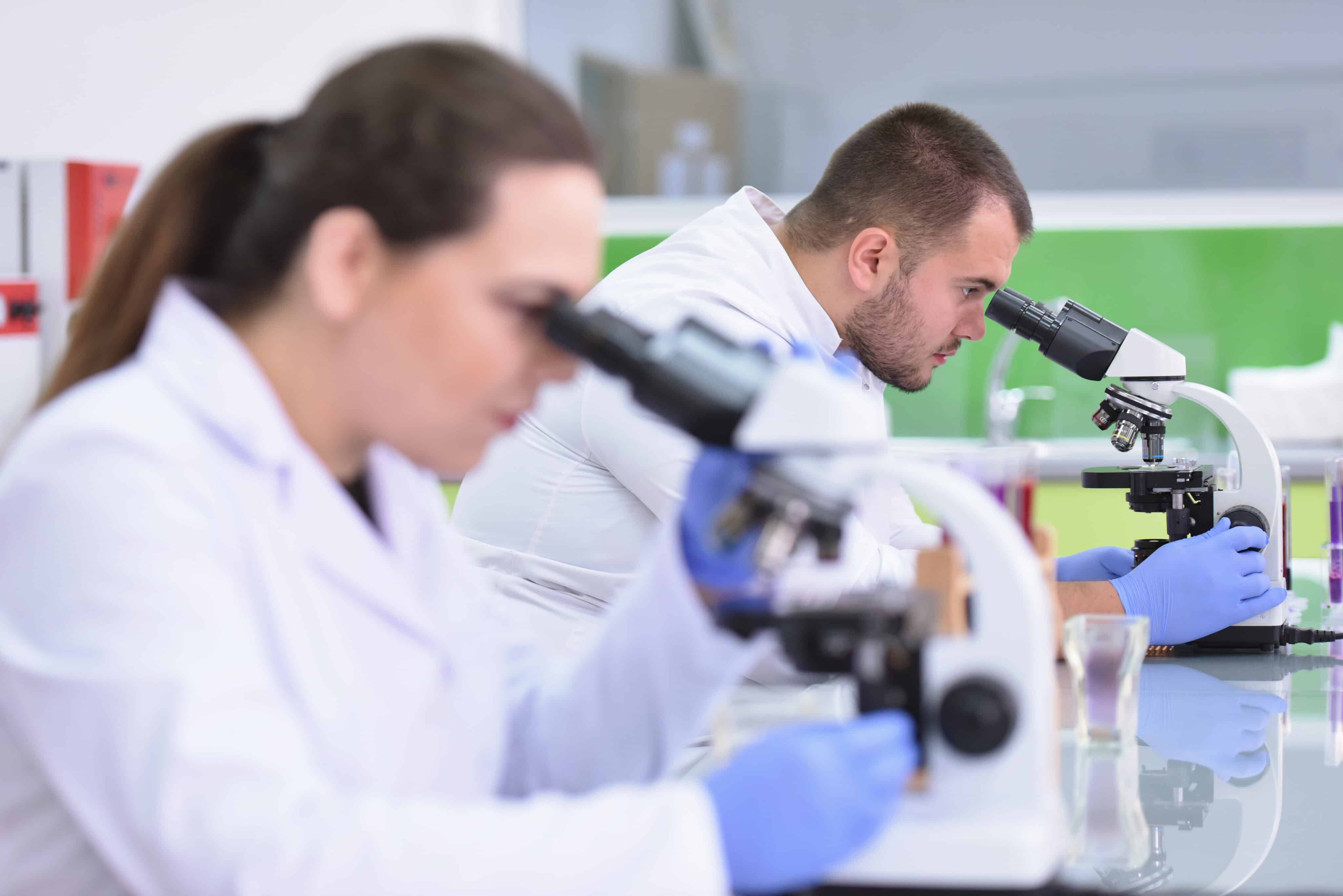
If you’re looking to familiarize yourself with the different parts of a microscope, you’re in luck! This article is about the diaphragm on microscopes, what it is, and how it works.
What Is a Diaphragm on a Microscope?
An iris diaphragm is an adjustable aperture used to control the amount of light that passes through the microscope objective. It is typically located in the microscope condenser lens system and consists of a series of overlapping blades that you can adjust to change the size of the aperture.
What Does the Diaphragm Do?
By controlling the size of the aperture, you can use the iris diaphragm to adjust the amount of light that reaches the specimen, improving image quality and contrast. This is particularly useful for specimens with different levels of transparency or reflectivity and samples that require high levels of illumination.
In addition to controlling the amount of light, you can also use the iris diaphragm to adjust the microscope’s depth of field. By adjusting the size of the aperture, the microscope operator can change the amount of light focused on a particular plane of the sample, which can affect the sharpness of the image and the depth of field.
Overall, the iris diaphragm is a valuable accessory for microscopes that can improve image quality and contrast by controlling the amount of light that reaches the sample and adjusting the depth of field of the microscope.
Understanding the Microscope Diaphragm
Let’s delve deeper into how the diaphragm works within the microscope.
As we explained above, the diaphragm on a microscope is an adjustable aperture located in the condenser lens system. Its primary function is to control the amount of light that passes through the specimen and reaches the objective lens.
The diaphragm consists of a series of overlapping blades that you can adjust to change the size of the aperture. By adjusting the size of the aperture, you can control the amount of light that reaches the specimen, which is essential for optimizing image quality and contrast.
When the aperture is wide open, a large amount of light reaches the specimen, which can result in a bright but blurry image. When the aperture is closed down, less light reaches the specimen, but the image becomes sharper and more contrasted. Adjusting the diaphragm allows the operator to find the optimal balance between brightness and sharpness for a given specimen.
You can also use the diaphragm to adjust the microscope’s depth of field. By adjusting the size of the aperture, the operator can change the amount of light focused on a particular plane of the specimen. This affects the image’s sharpness and the microscope’s depth of field.
The diaphragm on a microscope is an essential tool for controlling the amount of light that reaches the specimen, optimizing the image quality and contrast, and adjusting the depth of field of the microscope.
Common Uses of a Microscope Diaphragm
The diaphragm on a microscope is an important tool that helps to control the amount of light that reaches the specimen and optimize the image quality and contrast. There are several reasons why the diaphragm is an essential microscope part, reflected in the tasks it’s most commonly used for. These include:
Control of light intensity
By adjusting the size of the aperture, the diaphragm can control the amount of light that reaches the specimen. This is important because too much light can cause glare, wash out details, and reduce contrast, while too little light can make the image dim and hard to see. With the diaphragm, the operator can adjust the amount of light to obtain the best possible image.
Adjustment of the depth of field
By changing the size of the aperture, the diaphragm can also adjust the microscope’s depth of field. Depth of field refers to the distance between the nearest and farthest objects in a specimen that is in focus simultaneously. By controlling the amount of light that reaches the specimen, the diaphragm can adjust the depth of field, allowing the operator to focus on specific parts of the specimen and obtain the sharpest possible image.
Optimization of contrast
The diaphragm also helps to optimize the contrast of the image. By controlling the amount of light that reaches the specimen, the diaphragm can improve the image’s contrast, making it easier to see fine details and distinguish different parts of the specimen from each other.
Other Microscope Parts
A microscope is a complex instrument with several different parts. In addition to the diaphragm, other main parts of a microscope are:
- Eyepiece or ocular lens: This is the lens closest to the viewer’s eye and is used to magnify the image produced by the objective lens.
- Objective lens: This lens is closest to the specimen and magnifies the image. Microscopes typically have multiple objective lenses with varying magnifications.
- Nosepiece: This rotating component holds the objective lenses and allows the operator to switch between different magnification levels.
- Stage: This is the flat surface on which the specimen is placed for viewing. The stage may have clips or other mechanisms to hold the specimen in place.
- Illuminator: This light source provides the illumination needed to view the specimen. The illuminator may be built into the microscope or an external component.
- Condenser: This is the lens system that focuses the light from the illuminator onto the specimen.
- Diaphragm: This is an adjustable aperture in the condenser that controls the amount of light reaching the specimen.
- Coarse and fine focus controls: These are knobs or other mechanisms used to adjust the microscope’s focus. The coarse focus control makes larger adjustments, while the fine adjustment knob is used for more precise adjustments.
- Body tube: This is the component that connects the eyepiece to the objective lens and holds the other components of the microscope in place.
- Arm: This curved component connects the body tube to the microscope’s base and provides support and stability.
- Base: This is the bottom of the microscope and provides a stable platform for the other components.
In addition to these main components, microscopes may also have other features and accessories, such as filters, polarizers, and cameras, depending on the specific type and application of the microscope.
In Conclusion
In summary, a diaphragm is an essential tool that helps to optimize the image quality and contrast of a microscope by controlling the amount of light that reaches the specimen, adjusting the depth of field, and improving contrast. It allows operators to obtain the sharpest, most detailed images of specimens, critical for scientific research and diagnosis in medicine and biology. It’s a critical piece of the puzzle that is a microscope, helping scientists get the job done!

