Table of Contents
- What Is Darkfield Microscopy? Core Concept and Search Intent
- How Darkfield Illumination Works: Scattering, Numerical Aperture, and Stop Geometry
- When to Choose Darkfield vs Brightfield, Phase Contrast, and Fluorescence
- Visualizing Waterborne Microorganisms and Plankton for Education and Ecology
- Detecting Particulates for Cleanliness Inspection and Quality Control
- Trace Fibers, Hair, and Filaments in Forensic and Material Surveys
- Edges, Scratches, and Defects in Microelectronics and Polished Metals
- Practical Workflow and Sample Preparation for Robust Darkfield
- Quantitative Considerations: Exposure, Dynamic Range, and Background
- Troubleshooting Common Darkfield Artifacts
- Frequently Asked Questions
- Final Thoughts on Choosing the Right Darkfield Microscopy Approach
What Is Darkfield Microscopy? Core Concept and Search Intent
Darkfield microscopy is a contrast method that renders the background black while making tiny, light-scattering structures appear bright and high-contrast. Instead of transmitting direct light into the objective lens (as in brightfield), darkfield uses an oblique, hollow cone of illumination so that only light scattered by the specimen reaches the objective. The result is a visually striking view that highlights edges, fine particulates, and microorganisms that might otherwise be nearly invisible. When people search for darkfield microscopy, they are typically looking to understand how it differs from brightfield, what equipment it requires, and what kinds of samples it reveals best—ranging from waterborne microorganisms to particulate contamination on materials.

Attribution: Zephyris
This article focuses on real-world applications of darkfield microscopy across environmental, educational, and industrial contexts. You will learn the underlying physics that make darkfield work, how to compare it to other contrast methods, and how to apply it to specific tasks—such as surveying pond water for plankton or screening surfaces for dust and scratches. We will also cover practical setup guidance, quantitative imaging considerations, and troubleshooting tips. If you want a refresher on the core optical relationships that govern darkfield performance, jump to How Darkfield Illumination Works. If you are deciding whether darkfield is the right tool for your target sample, start at When to Choose Darkfield vs Other Methods.
How Darkfield Illumination Works: Scattering, Numerical Aperture, and Stop Geometry
At the heart of darkfield microscopy is a simple geometric idea: block the central, undeviated rays so that the objective lens only captures light that has been scattered by the specimen. The black background forms because direct illumination misses the objective’s front aperture. When a transparent or low-contrast structure is present, it scatters (re-directs) some of the oblique illumination into the objective, creating a bright signal against darkness.
Annular illumination and the role of the central stop
Darkfield requires either a dedicated darkfield condenser that generates a hollow cone of light or a patch stop (central stop) placed in the light path of a brightfield condenser. The central stop removes axial rays, producing an annular beam that strikes the specimen at high angles. The objective is positioned so that these high-angle, unscattered rays bypass it entirely. Only light that the specimen redirects (scatters) enters the objective and forms the image.
Numerical aperture (NA) relationships
For effective darkfield, the condenser’s numerical aperture (NA) must be set so that its illumination cone lies outside the objective’s NA. Stated qualitatively: the objective should not accept the direct illumination. A common practical guideline is that the inner NA of the illumination annulus should be larger than the objective’s NA so the direct rays miss the objective. In other words, the condenser should be adjusted (or selected) so that the background remains dark without a specimen in place. For higher NA objectives, this often necessitates a specialized darkfield condenser (sometimes oil-immersion) capable of delivering sufficiently oblique illumination.
This relationship ensures that the field stays dark in the absence of scattering, while even minute particles can produce bright signals. The mechanism does not increase the objective’s fundamental resolving power, which is governed by the objective NA and wavelength, but it can strongly improve contrast for small, scattering features. The Abbe diffraction limit (for incoherent imaging) can be expressed as resolution ≈ 0.61 · λ / NA_objective. Darkfield does not circumvent this limit; instead, it selectively enhances high-frequency content (edges, small objects) by suppressing low-frequency background.
Scattering regimes: Rayleigh to Mie
Scattering intensity depends on the size parameter (roughly the ratio of particle size to wavelength) and the refractive index contrast between specimen and medium. Extremely small particles (much smaller than the wavelength) follow Rayleigh-like scattering trends and may still be detectable in darkfield if the background is sufficiently clean and the illumination is optimized. Larger particles and edges fall into Mie-like or geometric scattering regimes, often producing pronounced bright rims and halos. Importantly, detected brightness in darkfield is not a simple measure of particle size or concentration; imaging conditions (illumination angle, NA, medium refractive index, and camera exposure) strongly influence appearance, as discussed in Quantitative Considerations.
Dry vs oil darkfield condensers and compatibility
Lower-magnification objectives often work well with a dry darkfield condenser or a central stop in a brightfield condenser since these objectives typically have modest NA values. As objective NA increases, the condenser must provide even higher-angle illumination; this is where an oil darkfield condenser can be required, because it supports higher effective NA in the illumination path. Always check practical alignment advice to maintain a dark background and avoid leakage of the direct beam into the objective.
Darkfield and resolution: contrast versus detail
Darkfield often reveals fine, filamentary structures and subvisible dust by boosting contrast, but it does not surpass the resolution defined by the objective and wavelength. The improvement you perceive is typically due to contrast gain for scattering features after low-frequency background has been suppressed by the stop. This is a powerful advantage for
applications like particulate inspection and microorganism surveys, where clear detection is more important than color rendering or absolute intensity accuracy.
When to Choose Darkfield vs Brightfield, Phase Contrast, and Fluorescence
Darkfield is one member of a family of contrast-enhancement modalities. Understanding its strengths relative to other methods helps you select the right tool for a given specimen and task.
Darkfield vs brightfield

Attribution: Zephyris
- Brightfield is versatile and straightforward, excelling with stained or intrinsically colored samples. Without staining, low-contrast transparent objects can be hard to see.
- Darkfield dramatically boosts visibility of edges, small particulates, and thin filaments by making the background black. It is often the easiest way to detect tiny contaminants or fast-moving, nearly transparent planktonic organisms.
- If color fidelity and tonal gradation are essential (e.g., histological stains), brightfield is typically preferred. For rapid screening of dust or microbes in a wet mount, darkfield can be superior.
Darkfield vs phase contrast
Attribution: Zephyris
- Phase contrast transforms phase variations into intensity differences, improving visibility of transparent specimens such as cells in culture. It requires phase-contrast objectives and corresponding phase annuli.
- Darkfield does not require phase-specific objectives, but it does require appropriate stops and condensers. It yields higher edge emphasis and suppresses background more strongly than phase contrast, which can be beneficial for detecting small particles.
- For detailed internal morphology of larger transparent cells, phase contrast often provides more informative tonal detail than darkfield. For fine debris, thin fibers, and small plankton, darkfield can be more sensitive.
Darkfield vs differential interference and oblique illumination
- Other techniques accentuate gradients or relief-like contrast and may require specialized optics. Darkfield is simpler to add to many stands via a stop or dedicated condenser.
- Oblique illumination (without a full darkfield stop) offers directional shadowing but may not fully eliminate background.
Darkfield vs fluorescence
- Fluorescence offers molecular specificity using labeled probes, at the cost of added complexity, filters, and photobleaching concerns.
- Darkfield is label-free and quick to deploy; it excels where intrinsic scattering contrast suffices (e.g., non-fluorescent particulates, unlabeled plankton).
- For targeted identification of a particular species or molecule, fluorescence is unmatched. For general surveys and cleanliness checks, darkfield is often faster and more economical.
Still deciding? The scenarios in Waterborne Microorganisms and Particulate Inspection illustrate where darkfield shines.
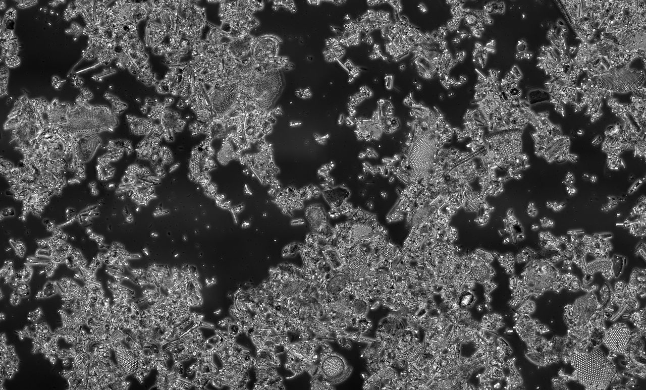
Visualizing Waterborne Microorganisms and Plankton for Education and Ecology
One of the most engaging uses of darkfield microscopy is the exploration of pond water, freshwater micro-ecosystems, and simple plankton samples. Many aquatic microorganisms are largely transparent and produce weak contrast in brightfield unless stained, which is impractical for live observation. Darkfield turns their edges and internal features into bright, sparkling structures against a black background, making it easier to see movement, morphology, and interactions in real time.

Attribution: KarlGaff, Art of Science Photography
What kinds of organisms look best in darkfield?
- Flagellates and ciliates: Fine appendages and ciliary beating produce flickering highlights, aiding recognition of locomotion patterns.
- Diatoms and algae: Siliceous frustules and cell walls often scatter strongly, revealing outlines and striations, especially at moderate magnifications.
- Rotifers and small crustaceans: Edges and feeding appendages stand out; darkfield reveals setae and spines that can be subtle in brightfield.
- Filamentous cyanobacteria and algae: Thin filaments become brightly traced lines, improving visibility in mixed communities.
- Protozoa with vacuoles or inclusions: Internal refractive structures create bright spots that move dynamically.
Why darkfield is appealing for live observation
- Label-free: No staining or fluorophores required; organisms remain undisturbed.
- Motion-friendly: High-contrast silhouettes against dark backgrounds help track motile species.
- Edge emphasis: Outlines, spines, and cilia are easy to spot, which aids qualitative identification in educational contexts.
Magnification and NA considerations for pond samples
For scanning large fields with many species, low to moderate magnification (e.g., 4×–20× objectives) balances field size and detail. Because the objective NA is relatively low in this range, achieving a dark background with a dry darkfield condenser or a central stop is usually straightforward. As you move to higher magnification to inspect fine details, the objective NA rises, so the condenser must deliver higher-angle illumination to preserve the dark background. If direct light leaks into the objective, the field will wash out. Revisit NA relationships to understand why this happens and how to correct it by adjusting stop alignment.
Interpreting appearance: halos, twinkling edges, and speckle
Bright rims or halos around objects are characteristic of darkfield. They arise from high-angle scattering at edges and refractive index discontinuities. Very small particles can display a
“twinkling” effect as they move in and out of focus, which helps distinguish them from fixed debris on the slide. However, halo width is not a direct size measurement. Remember that apparent brightness depends on multiple factors (size, shape, refractive index, illumination angle, and focus). For guidelines on interpreting intensity and avoiding overexposure, see Quantitative Considerations.
Educational use cases
- Surveying biodiversity: Rapidly locate diverse taxa and behavioral modes (swimmers, grazers, filter feeders) without staining.
- Food web demonstrations: Visualize predator-prey interactions and feeding currents; rotating the field of view helps learners track movement paths.
- Seasonal dynamics: Compare plankton communities across seasons or habitats to discuss ecological succession and environmental factors.
Darkfield’s ability to highlight motion and edges makes it ideal for classroom demonstrations and outreach events. When you need finer internal details or optical sectioning, you can complement darkfield with other methods discussed in contrast comparisons.
Detecting Particulates for Cleanliness Inspection and Quality Control
In manufacturing and materials handling, tiny particles can cause serious downstream problems—from optical scattering on lenses to shorts on microelectronic assemblies. Darkfield is widely used for cleanliness screening because it highlights minute debris on otherwise smooth surfaces. The black background intensifies contrast for dust, fibers, pits, and scratches, making darkfield a practical, label-free inspection tool during process control or incoming material assessment.
Why darkfield reveals particulates so effectively
- High-angle illumination accentuates discontinuities: Edges, steps, and surface asperities scatter strongly.
- Suppressed background reduces glare: With direct light excluded, specular reflections are minimized, so small particles pop out.
- Fine features appear as point-like or rim-brightened objects: Helpful for rapid detection against uniform or reflective substrates.
Typical targets in cleanliness checks
- Dust and fibers on optical components, wafers, or polished coupons.
- Residue and overspray on coated surfaces.
- Abrasive grains and wear particles in tribological tests.
- Contamination on filters, membranes, or swabs after sample collection.
Reflected-light darkfield for surfaces
While transmitted darkfield is suitable for thin or transparent samples, many cleanliness tasks use reflected-light darkfield to illuminate opaque surfaces at oblique angles. This can be accomplished with epi-illumination modules designed to deliver off-axis light that strikes the surface and misses the objective unless scattered. The principle mirrors transmitted darkfield: direct specular rays are kept out of the objective, while scattered light from particles and defects is collected. For practical setup, see Practical Workflow and Troubleshooting Artifacts.
Qualitative versus quantitative outcomes
Darkfield excels at qualitative detection—rapidly answering “Is something there?” It can also support semi-quantitative assessments, such as estimating relative cleanliness by counting bright events per field. However, absolute quantification of size and mass from darkfield intensity alone is not straightforward because scattering brightness depends on particle composition, shape, and the exact illumination geometry. If you require precise metrology, pair darkfield with calibrated imaging, or combine it with complementary modalities. We cover exposure and calibration concepts in Quantitative Considerations.
Trace Fibers, Hair, and Filaments in Forensic and Material Surveys
Darkfield is a powerful way to screen for trace fibers, hair, and filaments on adhesive lifts, garments, or environmental swabs in non-clinical, educational, and industrial contexts. Long, thin fibers that blend into brightfield backgrounds become prominent, glowing threads under darkfield. This makes preliminary surveys faster and reduces eye strain during prolonged inspections.

Attribution: Engjw
Advantages for filamentous targets
- Edge emphasis and haloing outline fine filaments even when they are colorless or translucent.
- Crossed or layered fibers remain distinguishable due to rim-brightening at each boundary.
- Crimp, diameter variation, and surface texture can be recognized qualitatively.
Substrate considerations
Because the background must remain dark, choose substrates with minimal scattering and glare when preparing educational demos. Transparent adhesive films can work well in transmitted darkfield if the adhesive is optically clean. For opaque lifts or garments, reflected-light darkfield is suitable; adjust illumination angle to suppress direct reflections. When in doubt, compare views across contrast methods to verify features are not artifacts.
Educational and outreach use
- Textile science demonstrations: Compare fiber morphologies across natural and synthetic materials in a visually compelling way.
- Environmental surveys: Screen for airborne fibers on sticky slides exposed in different rooms or outdoor settings.
- Introductory trace analysis: Teach the concept of screening versus confirmation, emphasizing that darkfield is a detection tool, not a definitive identifier.
Edges, Scratches, and Defects in Microelectronics and Polished Metals
Electronic assemblies, precision-machined parts, and polished metallographic samples benefit from darkfield’s knack for accentuating edges, micro-scratches, and pits. Even when a surface appears mirror-smooth in brightfield, darkfield can unveil subtle process marks that hint at fabrication issues or wear.
Reflected-light darkfield for patterned and polished surfaces
In microelectronics, a patterned surface includes abrupt changes in height and refractive index between features like traces, vias, and passivation layers. Oblique illumination scatters from these discontinuities, lighting them up against a dark substrate. For polished metals, faint scratches that run nearly parallel to the illumination direction may be subdued; rotating the sample or changing the azimuth of illumination can cause different scratches to light up. This rotation strategy is a practical technique also relevant to particulate inspections.
Complementary modalities
- Brightfield: Confirms color and general reflectivity.
- Oblique illumination without a full darkfield stop: Provides directional shadowing for depth cues.
- Interference-based surface profiling (outside the scope here): Useful for measuring step heights; darkfield can pre-screen regions of interest.
Interpreting scattered highlights
Under darkfield, sharp edges and pits often appear as intensely bright rims or points. The intensity depends on local slope and orientation relative to illumination. Consequently, bright spots indicate presence of a discontinuity but are not a direct measure of depth or height. For metrology, pair darkfield with calibrated imaging or different contrast techniques, while using darkfield as a rapid locator for areas needing closer analysis, as expanded in Quantitative Considerations.
Practical Workflow and Sample Preparation for Robust Darkfield
Small changes in alignment and cleanliness can make or break a darkfield image. While exact procedures vary by microscope model, the following general practices help deliver consistent, high-contrast results without turning this into a step-by-step laboratory protocol.
Maintain a truly dark background
- Center the stop: Ensure the central stop (or darkfield condenser annulus) is concentric with the optical axis so that direct light is excluded from the objective. Many stands allow you to inspect the back focal plane to verify alignment.
- Match NA: Select or adjust the condenser so that the inner NA of the illumination exceeds the objective’s NA, as described under NA relationships. If the background glows even without a sample, the stop is too small or misaligned.
- Control stray light: Close room lights and shield shiny surfaces near the microscope. Darkfield is sensitive to stray reflections.
Prepare clean, flat samples
- Use clean slides and coverslips to avoid spurious scatterers. Handle edges to minimize fingerprints and oils.
- For wet mounts (e.g., pond water), avoid thick bubbles and large debris clumps that can dominate scattering.
- For reflected-light darkfield, ensure the surface is free of smudges that produce broad glare; use gentle, appropriate cleaning methods suitable for the material.
Optimize focus and depth
- Darkfield images can be visually rich but shallow in useful depth due to the oblique illumination. Use fine focus to scan through layers, especially with thick or three-dimensional samples.
- For particulate inspection on flat surfaces, ensure the focal plane coincides with the surface to avoid misjudging size or location.
Camera and viewing comfort
- Darkfield is often dimmer than brightfield because most illumination is excluded. Increase exposure time moderately, use higher sensor gain thoughtfully, and stabilize the setup to reduce motion blur. More on this in Quantitative Considerations.
- Use a comfortable viewing brightness through eyepieces; avoid excessive light levels that raise background spill or cause eye fatigue.
Rheinberg and color contrast variants

Attribution: Albega (original uploader); derivative work: Dietzel65
Some practitioners enjoy Rheinberg illumination, where colored filters in the stop and annulus yield aesthetic contrasts (e.g., blue background, red specimen). While visually compelling for outreach, the same NA and alignment principles apply. Keep in mind that colorized backgrounds are not quantitative.
Quantitative Considerations: Exposure, Dynamic Range, and Background
Because darkfield strongly suppresses the background, small signals from scattering objects can span a narrow range of intensities. Optimizing exposure and dynamic range is crucial for documentation, comparison across samples, and minimizing artifacts.
Exposure control and saturation
- Avoid saturating bright edges: Once a pixel saturates, you lose information about intensity variations and halo structure. Set exposure so the brightest highlights remain below clipping.
- Beware of dim backgrounds creeping up: If you increase exposure too much, small amounts of stray light or internal reflections will lift the background from black to gray, reducing contrast.
- Stabilize the sample: For live plankton, choose exposure times short enough to avoid motion blur, balanced against noise. If the scene is too dim, consider slightly higher gain or brighter illumination rather than excessive exposure.
White balance and color rendition
Darkfield is primarily about intensity contrast, so color fidelity is less critical than in brightfield histology. Nevertheless, calibrating white balance ensures consistent documentation when comparing samples or teaching. For reflected-light darkfield, metallic surfaces can bias color via spectral reflectivity; setting a custom white balance on a clean, uniform region can help.
Measuring size and counting particles
- Calibrate pixel size: Use a stage micrometer to map pixels to micrometers at each magnification. This allows direct size measurement of particles and features.
- Segment with caution: Automated thresholding can be effective because the background is dark, but haloing may overestimate object size. Consider edge-aware methods to delineate true particle boundaries.
- Avoid using brightness as a proxy for mass: Scattering intensity depends on more than size (shape, refractive index, and angle matter). Use geometric measurement rather than intensity summation for comparisons.
Resolution and spatial frequency content
Darkfield filters out low spatial frequencies, emphasizing edges and fine details. This has two implications: (1) images appear crisper for filamentary and particulate features, and (2) smooth intensity gradients can become difficult to interpret. Remember that the 0.61 · λ / NA_objective relationship still sets the fundamental limit for resolving two features. Oblique illumination can shift the distribution of detectable frequencies, but it does not grant resolution beyond what the objective NA supports.
Troubleshooting Common Darkfield Artifacts
Darkfield’s beauty lies in its sensitivity—but this also means it readily reveals unwanted scatterers and alignment errors. Here are practical ways to detect and correct common issues.
Glowing background or uneven field
- Cause: Stop misalignment or undersized stop allows direct light into the objective.
- Fix: Re-center the stop, verify condenser iris and annulus position, and check that the condenser NA is set so the inner annulus exceeds the objective NA. See NA relationships.
Hot spots and flare
- Cause: Stray reflections from dust on optics, fingerprints, or shiny slide edges.
- Fix: Clean accessible optical surfaces with appropriate methods; trim or mask shiny edges; reduce ambient light.
Halos that obscure detail
- Cause: Overexposure exaggerates halos; out-of-focus layers contribute bright blur.
- Fix: Reduce exposure until highlights are controlled; refocus in small steps and consider thinner samples or careful mounting.
Particles that vanish when refocusing
- Cause: Debris on the coverslip or condenser rather than in the specimen plane.
- Fix: Move the slide: true specimen particles will shift relative to fixed debris; clean slide surfaces.
Apparent double edges
- Cause: Coherent-like interference fringes or scattering from layered structures viewed with oblique illumination.
- Fix: Slightly alter illumination angle or rotate the sample; confirm with an alternative contrast method as a control.
Frequently Asked Questions
Does darkfield microscopy increase resolution beyond brightfield?
No. The fundamental resolution limit for incoherent imaging is governed by the objective’s numerical aperture and the wavelength of light, typically approximated by 0.61 · λ / NA_objective. Darkfield improves contrast by suppressing the background and emphasizing scattered light from edges and small particulates, but it does not surpass the objective’s resolution. In some cases, oblique illumination redistributes spatial frequencies, changing the appearance of fine structure, yet the underlying resolution bound remains set by the objective NA.
When should I choose transmitted vs reflected darkfield?
Use transmitted darkfield for transparent or thin specimens—such as pond water, thin films on glass, or particles collected on transparent membranes—where light passes through the sample. Choose reflected darkfield for opaque surfaces—like machined parts, polished metals, or microelectronic assemblies—where illumination must strike and scatter from the surface. In each case, the goal is the same: exclude direct rays from the objective so only scattered light is collected. For more on setup considerations, see Practical Workflow.
Final Thoughts on Choosing the Right Darkfield Microscopy Approach
Darkfield microscopy remains one of the most accessible, visually compelling ways to reveal what is otherwise hidden at the microscopic scale. By blocking direct illumination and capturing only scattered light, it excels at detecting microorganisms, particulates, fibers, and surface defects across educational, environmental, and industrial contexts. It is fast, label-free, and complementary to brightfield, phase contrast, and other contrast modalities.
To achieve reliable results, remember three essentials: (1) align the stop so the background truly goes black, (2) match condenser and objective NAs so direct rays miss the objective, and (3) optimize exposure to preserve highlight detail without lifting the background. When you need to measure sizes or compare samples quantitatively, calibrate your imaging scale and avoid using brightness as a proxy for mass or composition, as outlined in Quantitative Considerations. For mixed tasks—such as scanning for debris and then documenting morphology—pair darkfield with a secondary method described in contrast comparisons.
Whether you are surveying pond life, checking a lens for dust, or highlighting trace fibers on a substrate, darkfield can provide a decisive first look. Explore more microscopy topics and practical guides in future installments, and consider subscribing to our newsletter to receive upcoming articles on contrast methods, imaging workflows, and application-focused tutorials.

